Tag Archives: lungs specialist in karimnagar

Pediatric Bronchoscopy: Purpose, Procedure & Recovery Guide

Understanding Pediatric Bronchoscopy: Purpose, Procedure, and Recovery

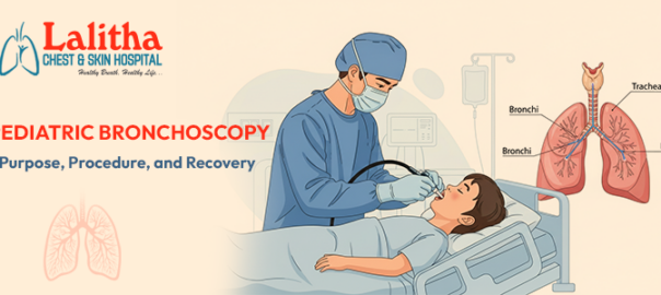
Pediatric bronchoscopy is a specialized diagnostic and therapeutic procedure used to examine a child’s airways, including the throat, windpipe, and lungs. It allows doctors to identify and treat various respiratory problems that may not be visible through regular imaging tests like X-rays or CT scans. This minimally invasive procedure plays a crucial role in understanding the cause of persistent cough, breathing difficulties, or recurrent lung infections in children.

With expert insights from Dr. Raj Kumar Korra at Lalitha Chest and Skin Hospital, this blog explores the purpose, procedure, and recovery process in detail.

What Is Pediatric Bronchoscopy?

“A bronchoscopy involves inserting a thin, flexible tube called a bronchoscope through the nose or mouth into the lungs. The tube has a light and a camera that help the pulmonologist clearly view the airways. In children, this procedure is performed by a pediatric pulmonologist with expertise in handling delicate airways and ensuring maximum safety and comfort,” explains Dr. Raj Kumar Korra, MD, the best pulmonologist in Karimnagar.

Depending on the case, doctors may use:

  • Flexible bronchoscopy: Commonly used for diagnosis and minor treatments.
  • Rigid bronchoscopy: Used for removing foreign objects or treating major airway blockages.

Purpose of Pediatric Bronchoscopy:

Doctors recommend bronchoscopy for children when symptoms or imaging tests suggest airway or lung problems. Some common reasons include:

  1. Persistent cough or wheezing
  2. Recurrent lung infections
  3. Difficulty breathing or noisy breathing
  4. Unexplained chest X-ray finding
  5. Suspected inhalation of a foreign object (like food or a small toy)
  6. Evaluation of airway abnormalities or structural defects
  7. Collection of mucus or tissue samples for laboratory testing

By directly visualizing the airways, the doctor can make an accurate diagnosis and plan the most effective treatment.

How the Procedure Is Performed:

Before the procedure, the child may be given sedation or general anesthesia to ensure they are comfortable and relaxed. During bronchoscopy:

  1. The doctor carefully guides the bronchoscope through the child’s nose or mouth into the airways.
  2. The camera provides a clear view of the airways on a monitor.
  3. The doctor may take mucus or tissue samples (biopsy) if needed.
  4. If any blockages or foreign bodies are found, they can often be removed during the same procedure.

The entire process typically takes 20 to 45 minutes, depending on the complexity.

Post-Procedure Care and Recovery

After the bronchoscopy, the child is monitored until the effects of anesthesia wear off. Mild throat discomfort, coughing, or hoarseness is common and typically resolves within one to two days.

Parents are advised to:

  • Offer soft foods and plenty of fluids once the child can swallow comfortably.
  • Avoid strenuous activity for the rest of the day.
  • Follow the doctor’s instructions regarding medications or follow-up visits.

If your child experiences persistent breathing difficulty, high fever, or severe chest pain, contact your healthcare provider immediately.

Is Pediatric Bronchoscopy Safe?

Yes, pediatric bronchoscopy is generally safe when performed by experienced specialists. Modern equipment and advanced anesthesia techniques have made the procedure low-risk. Complications such as infection or bleeding are rare and are carefully managed by the medical team.

Pediatric bronchoscopy is a valuable diagnostic tool for identifying and treating various respiratory conditions in children. It helps doctors visualize the airways directly, leading to accurate diagnoses and effective care. With skilled pediatric pulmonologists and proper post-procedure care, most children recover quickly and comfortably.

If your child has ongoing breathing issues or unexplained respiratory symptoms, consult a pediatric pulmonologist to determine if bronchoscopy might help identify the underlying cause.

If you’re in Karimnagar and searching for terms like “bronchoscopy for lung cancer,” “bronchoscopy treatment,” or “lung cancer bronchoscopy biopsy,” visit Lalitha Chest and Skin Hospital. Dr. Raj Kumar Korra, a leading lung specialist in Karimnagar, offers advanced and precise lung care. Book your appointment today by visiting our website: https://lalithachestandskinhospital.com/

What to Expect During Pleural Fluid Aspiration | Patient Guide

A Patient’s Guide to Pleural Fluid Aspiration: Diagnosis and Treatment Insights

Pleural fluid aspiration, also known as thoracentesis, is a common medical procedure that helps doctors diagnose and treat conditions affecting the lungs and the pleural space — the thin area between the lungs and chest wall. If your doctor has recommended this procedure, understanding what it involves can help ease any worries and prepare you for a smooth experience.

What is Pleural Fluid Aspiration?

The lungs are surrounded by two layers of tissue called the pleura, and between them is a small amount of fluid that allows smooth lung movement during breathing. However, due to infections, heart failure, lung disease, or other conditions, excess fluid can build up in this space — a condition known as pleural effusion, ” says Dr. Raj Kumar Korra, MD, the best pulmonologist in Karimnagar.

Pleural fluid aspiration is a procedure performed to remove some of this fluid for diagnostic or therapeutic purposes:

  • Diagnostic aspiration: Helps determine the cause of the fluid buildup.
  • Therapeutic aspiration: Helps relieve symptoms such as shortness of breath or chest discomfort.

With expert insights from Dr. Raj Kumar Korra at Lalitha Chest and Skin Hospital, this blog explores what pleural fluid aspiration is, why it is necessary, the procedure involved, post-procedure care, and diagnostic methods. Read on to learn more:

Why Is Pleural Fluid Aspiration Needed?

Your doctor may recommend this procedure if you experience symptoms like:

  • Persistent shortness of breath
  • Chest pain that worsens with deep breaths
  • Cough or heaviness in the chest
  • Abnormal chest X-ray or ultrasound findings

Common conditions that may cause pleural effusion include:

  • Pneumonia or lung infections
  • Heart failure
  • Liver or kidney disease
  • Tuberculosis (TB)
  • Lung cancer or metastatic disease

How Is the Procedure Performed?

Pleural fluid aspiration is generally a safe and minimally invasive procedure performed under local anesthesia. Here’s what you can expect:

  1. Preparation: You may be asked to sit upright and lean slightly forward. The doctor will clean the area on your back or side with an antiseptic solution.
  2. Anesthesia: Local anesthesia is given to numb the area, ensuring minimal discomfort.
  3. Fluid Removal: A thin needle or catheter is carefully inserted into the pleural space. The doctor withdraws a small amount of fluid for testing or removes excess fluid to relieve pressure.
  4. Post-procedure care: Once the needle is removed, the area is bandaged. You may be asked to undergo a chest X-ray to ensure there are no complications like air leaks.

What Happens to the Fluid Sample?

The fluid collected during the procedure is sent to a laboratory for detailed analysis. Tests may include:

  1. Cytology: To detect cancer cells.
  2. Microbiology: To identify infections such as tuberculosis or bacterial pneumonia.
  3. Biochemical analysis: To assess protein, glucose, and enzyme levels

These results help your doctor determine the underlying cause of the pleural effusion and guide appropriate treatment.

After the Procedure: What to Expect:

Most patients recover quickly after pleural fluid aspiration. Mild soreness or slight discomfort at the needle site is normal and should resolve within a day or two. Your doctor will give you specific aftercare instructions, such as:

  • Avoiding strenuous activities for 24 hours
  • Watching for symptoms like chest pain, shortness of breath, or fever
  • Keeping the bandage area clean and dry

If you experience sudden breathlessness or severe chest pain, contact your healthcare provider immediately.

Risks and Complications:

While pleural fluid aspiration is a safe procedure, some rare complications can occur, such as:

  • Pneumothorax (air leak in the lung)
  • Infection at the puncture site
  • Bleeding
  • Re-accumulation of fluid

Your doctor will take all precautions to minimize these risks using ultrasound guidance and sterile techniques.

Treatment After Diagnosis:

Once the cause of pleural effusion is determined, treatment focuses on managing the underlying condition.

For example:

  • Antibiotics for infections
  • Diuretics for heart failure
  • Anti-tuberculosis medications for TB
  • Oncology treatment for cancer-related effusions

In recurrent cases, procedures like pleurodesis (to prevent fluid from returning) may be recommended.

Pleural fluid aspiration is an important diagnostic and therapeutic procedure that provides vital information about lung and chest health. With skilled medical supervision, it offers quick relief from breathing discomfort and plays a key role in identifying the cause of pleural effusion.

If you have symptoms such as persistent breathlessness or chest heaviness, don’t ignore them. Consult a chest specialist for an accurate diagnosis and proper treatment plan.

If you’re in Karimnagar and searching online for terms like “pleural effusion lung cancer treatment,” “removing fluid from lungs surgery,”  or “lung pleural effusion treatment,” let us save you time by helping you book an appointment quickly and easily.

At Lalitha Chest and Skin Hospital, our team, led by Dr. Raj Kumar Korra, a lung specialist in Karimnagar, specializes in diagnosing and managing various lung conditions, including pleural effusion. We provide safe, precise, and patient-centered care using advanced diagnostic tools and expert medical support. Please visit our website to schedule your appointment today: https://lalithachestandskinhospital.com/

Bronchiectasis: Symptoms, Diagnosis, & Treatment Options

Bronchiectasis Awareness: Identifying Symptoms and Exploring Treatment Options

Bronchiectasis is a chronic condition that affects the lungs, leading to the abnormal and permanent widening of the airways. This widening impairs the lungs’ ability to clear mucus, which can lead to infections and further lung damage. Awareness and early diagnosis are crucial for managing the condition effectively.

“Bronchiectasis occurs when the bronchial tubes, which carry air to and from the lungs, become damaged and dilated, often due to infections or other medical conditions that inflame and injure the airways. This results in widened airways that are unable to clear mucus effectively, leading to a cycle of infection and inflammation,” says Dr. Raj Kumar Korra, MD, a lung specialist in Karimnagar.

With insights from Dr. Raj Kumar Korra at Lalitha Chest and Skin Hospital, this blog provides comprehensive information on bronchiectasis. We explore its symptoms, available treatment options, and effective management tips. Read on to learn more:

Common Symptoms of Bronchiectasis:

Recognizing the symptoms of bronchiectasis early can help in seeking timely medical intervention. Common symptoms include:

  1. Chronic Cough: A persistent cough that produces large amounts of mucus or phlegm.
  2. Sputum Production: Mucus may be thick, discolored, or even contain blood.
  3. Shortness of Breath: Difficulty breathing or feeling breathless, particularly during physical activities.
  4. Recurrent Respiratory Infections: Frequent lung infections or exacerbations that don’t resolve quickly.
  5. Wheezing: A whistling sound when breathing, often heard with a stethoscope.
  6. Fatigue: Persistent tiredness or feeling generally unwell.

If you experience these symptoms, it’s important to consult a healthcare professional for an accurate diagnosis and appropriate treatment plan.

What Causes and Risk Factors?

Bronchiectasis can result from various conditions and factors, including:

  1. Chronic Respiratory Infections: Repeated infections can damage the airways and lead to bronchiectasis.
  2. Cystic Fibrosis: A genetic disorder that causes thick, sticky mucus to build up in the lungs.
  3. Autoimmune Diseases: Conditions like rheumatoid arthritis can affect the lungs and lead to bronchiectasis.
  4. Bronchial Obstruction: Tumors or foreign objects blocking the airways can contribute to the development of bronchiectasis.
  5. Genetic Disorders: Conditions like Alpha-1 Antitrypsin Deficiency can increase the risk.

 Diagnosing Bronchiectasis:

Diagnosing bronchiectasis typically involves several steps:

  1. Medical History and Physical Examination: Your doctor will review your symptoms, medical history, and perform a physical exam.
  2. Imaging Tests: A chest X-ray or high-resolution computed tomography (HRCT) scan can reveal the characteristic widening of the airways.
  3. Pulmonary Function Tests: These tests measure how well your lungs are working and help determine the extent of airway damage.
  4. Sputum Culture: Analyzing mucus samples can identify any bacterial infections and guide treatment.
  5. CT Scan: Provides more detailed images of the lungs if needed.

Treatment for Bronchiectasis:

Although bronchiectasis cannot be cured, various treatment options are available to manage symptoms and enhance quality of life. Treatment generally focuses on:

  1. Medication
  • Antibiotics: Used to treat and prevent infections.
  • Bronchodilators: Help open the airways and improve breathing.
  • Mucolytics: Medications that thin mucus, making it easier to clear from the lungs.
  1. Airway Clearance Techniques
  • Chest Physiotherapy: Techniques like postural drainage and chest percussion help remove mucus from the airways.
  • Positive Expiratory Pressure (PEP) Therapy: Devices that help keep the airways open and promote mucus clearance.
  1. Lifestyle and Home Remedies
  • Hydration: Drinking plenty of fluids helps thin mucus, making it easier to expel.
  • Healthy Diet: A balanced diet can support overall health and immune function.
  • Regular Exercise: Helps improve lung function and overall well-being.
  1. Surgery

In severe cases, surgery may be considered to remove damaged sections of the lungs or to address specific complications.

 Living with Bronchiectasis:

Managing bronchiectasis requires a combination of medical treatment, lifestyle adjustments, and ongoing monitoring. Regular follow-ups with your healthcare provider are essential to track your condition and adjust treatments as needed.

Tips for Coping:

  • Stay Informed: Educate yourself about bronchiectasis and stay updated on new treatments and management strategies.
  • Join Support Groups: Connecting with others who have bronchiectasis can provide emotional support and practical advice.
  • Monitor Your Health: Keep track of your symptoms and report any changes to your healthcare provider promptly.

Bronchiectasis is a manageable condition with the right approach and treatment plan. By recognizing the symptoms early and exploring available treatment options, individuals with bronchiectasis can lead a fulfilling life. If you suspect you have bronchiectasis or are experiencing related symptoms, seek medical advice to receive a proper diagnosis and personalized care.

If you’re in Karimnagar and searching online for terms like “bronchiectasis treatment,” “lungs hospital near me,” or “best chest physician near me,” let us save you time by recommending Dr. Raj Kumar Korra at Lalitha Chest and Skin Hospital for expert care.

Lalitha Chest and Skin Hospital is Karimnagar’s top choice for bronchiectasis treatment, boasting an outstanding record in handling all cases. Led by Dr. Raj Kumar Korra, a renowned breathing specialist, we offer comprehensive care for lung conditions. For appointments, please visit our website: https://lalithachestandskinhospital.com/